How can we visualize a plant from within to diagnose the plant's health without cutting into its tissue? The solution can be found in ultra-high field MRI scanners; two to nine times stronger than hospital MRI scanners.
Author: Julia Krug, Laboratory at BioNanoTechnology, Wageningen University & Research
Magnetic Resonance Imaging (MRI) is a technique well-known in the medical field for diagnostics, as it can yield images without the need for an investigative surgery. These same capabilities can be used to image other specimen non-destructively, such as plants.
MRI is a relatively insensitive technique leading to low image resolutions. For small plant parts, such as flower stems, higher resolutions are needed. To circumvent this problem, stronger magnets can be used to obtain images at high spatial resolutions. In our research, we have access to ultra-high field scanners, such as a 14.1T system, which are between two to nine times stronger than hospital MRI scanners.
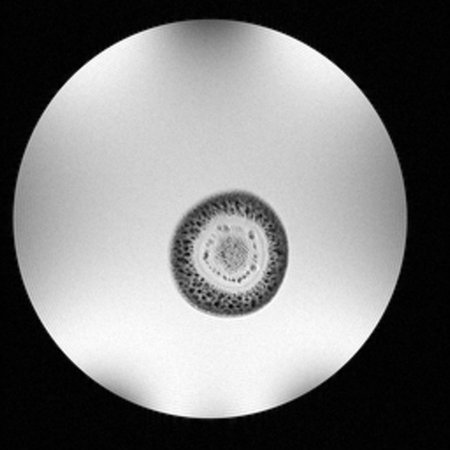
To illustrate the potential for MRI on plant specimens, in the figure we show a cross-section of a flower stem of 2.5 mm in diameter of a Lisianthus flower (Eustoma russelianum) taken at 14.1 T by Maurits Naber. In these images, we can distinguish the different tissues (xylem, phloem, pith) and potentially use them to diagnose plant health.

A high-resolution MRI image of a flower stem with an image resolution of 39 x 39 μm and a slice thickness of 1 mm.
At the Laboratory of BioNanoTechnology in Wageningen, we are investigating plant structures using ultra-high field MRI equipment. Maurits Naber has dedicated his BSc-thesis to this project and Dani Kortekaas will continue with this research during her MSc-thesis.